Home

Cura compensazione terreno di gioco lysotracker deep red Senza greca azienda agricola

LysoTracker™ Deep Red

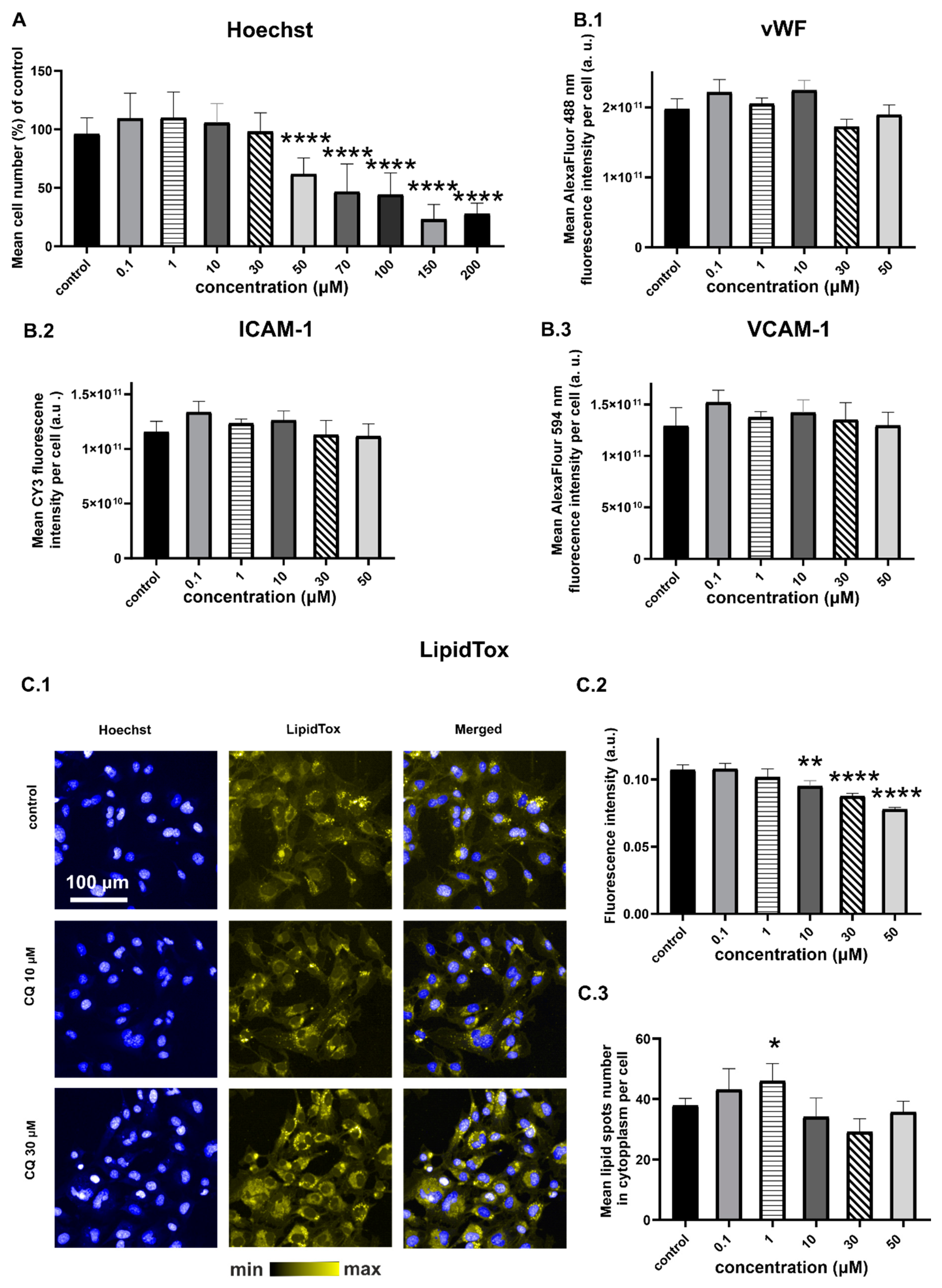
IJMS | Free Full-Text | Chloroquine-Induced Accumulation of Autophagosomes  and Lipids in the Endothelium
IJMS | Free Full-Text | Chloroquine-Induced Accumulation of Autophagosomes and Lipids in the Endothelium

STING signalling is terminated through ESCRT-dependent microautophagy of  vesicles originating from recycling endosomes | Nature Cell Biology
STING signalling is terminated through ESCRT-dependent microautophagy of vesicles originating from recycling endosomes | Nature Cell Biology

Concomitant evaluation of ER tracker and LysoTracker Deep Red (LTDR)... |  Download Scientific Diagram
Concomitant evaluation of ER tracker and LysoTracker Deep Red (LTDR)... | Download Scientific Diagram

a–c) Microscopy images of A549 cells stained by 20 × 10⁻⁶ m COE‐BBT for...  | Download Scientific Diagram
a–c) Microscopy images of A549 cells stained by 20 × 10⁻⁶ m COE‐BBT for... | Download Scientific Diagram

Cell-permeable organic fluorescent probes for live-cell long-term  super-resolution imaging reveal lysosome-mitochondrion interactions |  Nature Communications
Cell-permeable organic fluorescent probes for live-cell long-term super-resolution imaging reveal lysosome-mitochondrion interactions | Nature Communications

LysoTracker assay. LysoTracker red dye staining shows the distribution... |  Download Scientific Diagram
LysoTracker assay. LysoTracker red dye staining shows the distribution... | Download Scientific Diagram

Concomitant evaluation of ER tracker and LysoTracker Deep Red (LTDR)... |  Download Scientific Diagram
Concomitant evaluation of ER tracker and LysoTracker Deep Red (LTDR)... | Download Scientific Diagram

LysoBrite™ Deep Red | AAT Bioquest

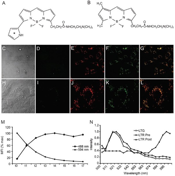
Molecules | Free Full-Text | Bioimaging of Lysosomes with a BODIPY  pH-Dependent Fluorescent Probe
Molecules | Free Full-Text | Bioimaging of Lysosomes with a BODIPY pH-Dependent Fluorescent Probe

Co-localization of C-1330 and LysoTracker red in lysosomes in A549 and... |  Download Scientific Diagram
Co-localization of C-1330 and LysoTracker red in lysosomes in A549 and... | Download Scientific Diagram

Deep-red to near-infrared fluorescent dyes: Synthesis, photophysical  properties, and application in cell imaging - ScienceDirect
Deep-red to near-infrared fluorescent dyes: Synthesis, photophysical properties, and application in cell imaging - ScienceDirect

Subcellular distribution of LysoTracker Deep Red (blue color), and... |  Download Scientific Diagram
Subcellular distribution of LysoTracker Deep Red (blue color), and... | Download Scientific Diagram

Photoconversion of Lysotracker Red to a green fluorescent molecule | Cell  Research
Photoconversion of Lysotracker Red to a green fluorescent molecule | Cell Research

70058-T | LysoView™ 633, trial size Quimigen

IJMS | Free Full-Text | Chloroquine-Induced Accumulation of Autophagosomes  and Lipids in the Endothelium
IJMS | Free Full-Text | Chloroquine-Induced Accumulation of Autophagosomes and Lipids in the Endothelium

FIRE-pHLy localizes to lysosomal compartments. (A−E) Representative... |  Download Scientific Diagram
FIRE-pHLy localizes to lysosomal compartments. (A−E) Representative... | Download Scientific Diagram

LysoTracker and MitoTracker Red are transport substrates of P‐glycoprotein:  implications for anticancer drug design evading multidrug resistance -  Zhitomirsky - 2018 - Journal of Cellular and Molecular Medicine - Wiley  Online Library
LysoTracker and MitoTracker Red are transport substrates of P‐glycoprotein: implications for anticancer drug design evading multidrug resistance - Zhitomirsky - 2018 - Journal of Cellular and Molecular Medicine - Wiley Online Library

LysoTracker™ Deep Red

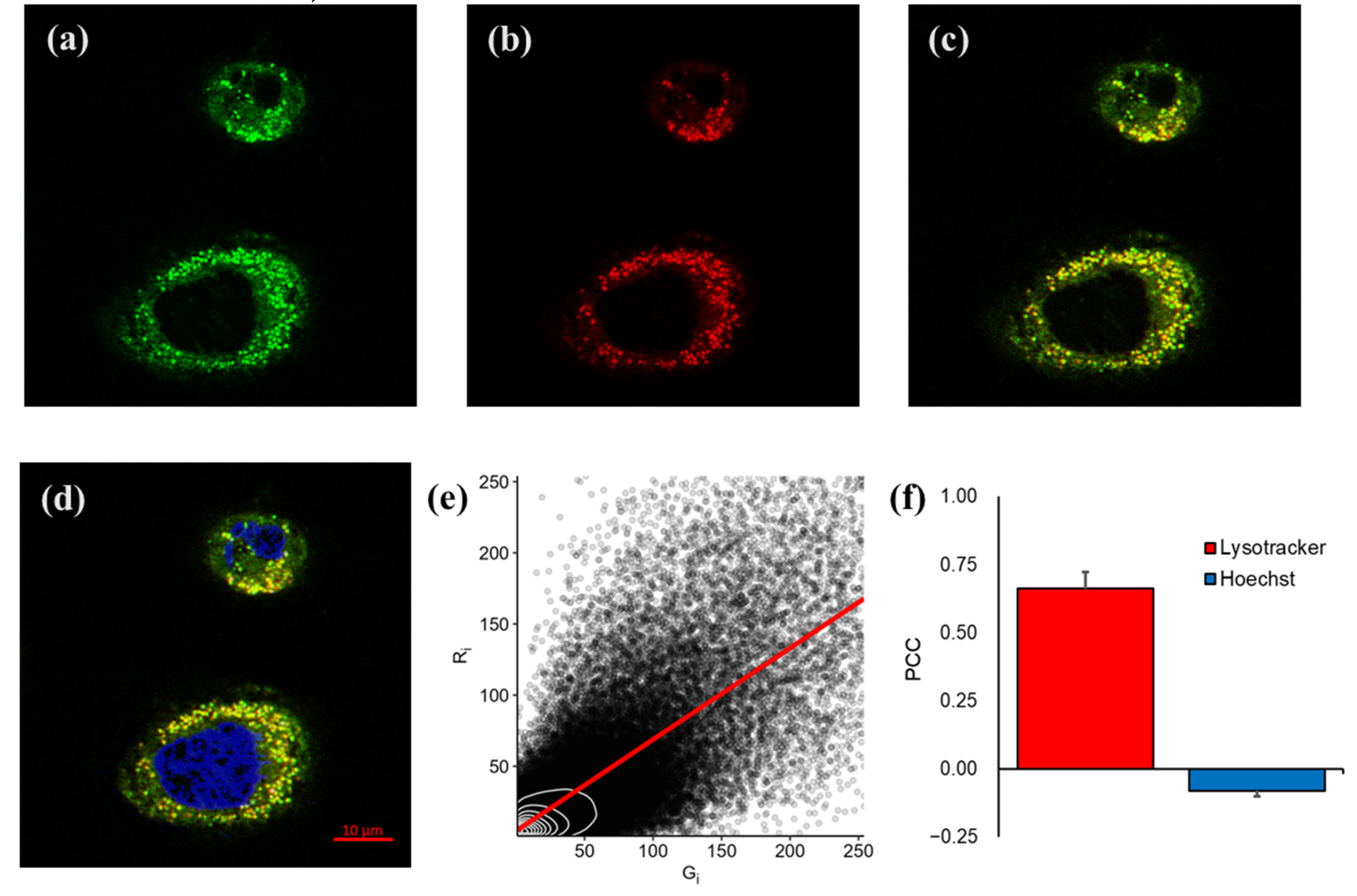
Cellular distribution and co-localization of compounds 11-13.C onfocal... |  Download Scientific Diagram
Cellular distribution and co-localization of compounds 11-13.C onfocal... | Download Scientific Diagram

Labeling and Visualization of Lysosome using Lysotracker

Protocol for evaluating autophagy using LysoTracker staining in the  epithelial follicle stem cells of the Drosophila ovary - ScienceDirect
Protocol for evaluating autophagy using LysoTracker staining in the epithelial follicle stem cells of the Drosophila ovary - ScienceDirect
LysoTracker™ Red DND-99, special packaging

LysoTracker™ Deep Red

Lysosomes - Flow Cytometry Core Facility

LysoTracker™ Deep Red

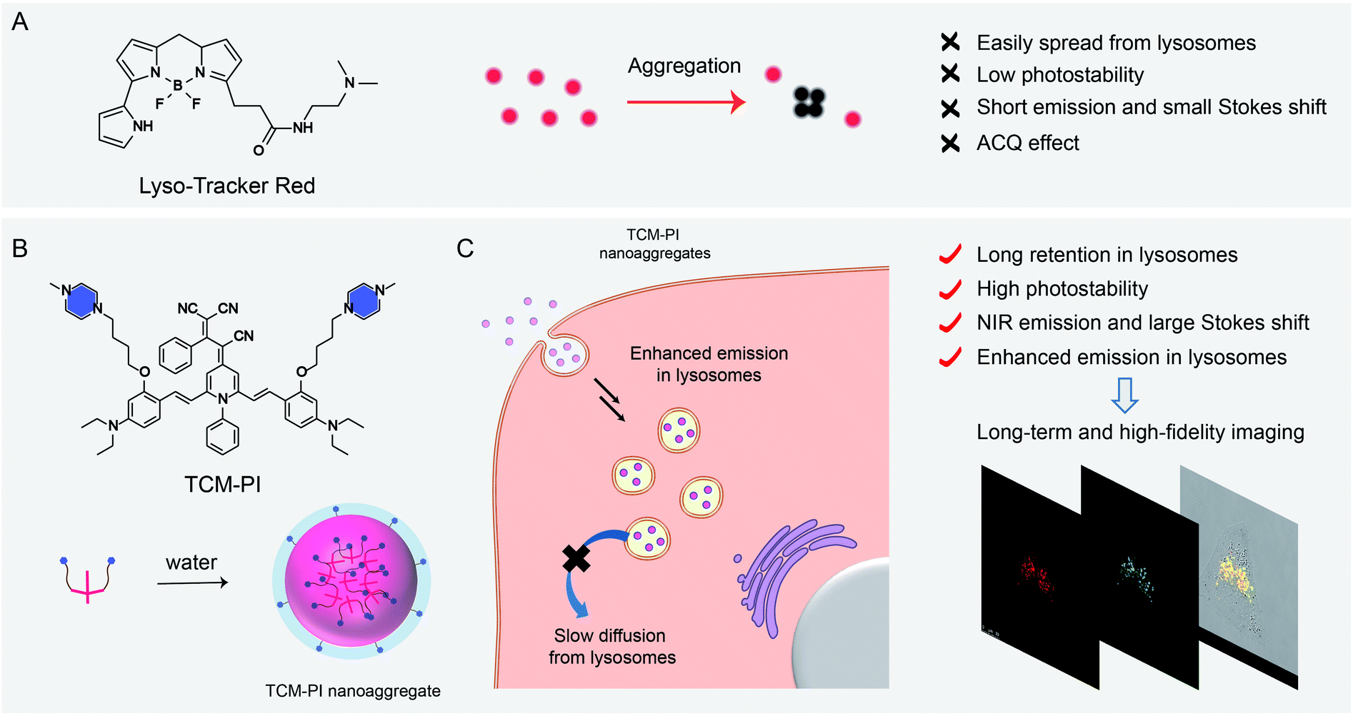
AIE-based nanoaggregate tracker: high-fidelity visualization of lysosomal  movement and drug-escaping processes - Chemical Science (RSC Publishing)  DOI:10.1039/D0SC04156D
AIE-based nanoaggregate tracker: high-fidelity visualization of lysosomal movement and drug-escaping processes - Chemical Science (RSC Publishing) DOI:10.1039/D0SC04156D

Use of LysoTracker to Detect Programmed Cell Death in Embryos and  Differentiating Embryonic Stem Cells | Protocol
Use of LysoTracker to Detect Programmed Cell Death in Embryos and Differentiating Embryonic Stem Cells | Protocol